Review Another Set of Terms Associated With a Skeletal Muscle Fiber/cell and the Sarcomere
Chapter 19. The Musculoskeletal System
19.four Musculus Wrinkle and Locomotion
Learning Objectives
By the terminate of this section, you will exist able to:
- Classify the different types of muscle tissue
- Explain the role of muscles in locomotion
Muscle cells are specialized for contraction. Muscles let for motions such as walking, and they also facilitate bodily processes such as respiration and digestion. The trunk contains three types of muscle tissue: skeletal musculus, cardiac muscle, and smooth musculus (Figure 19.33).
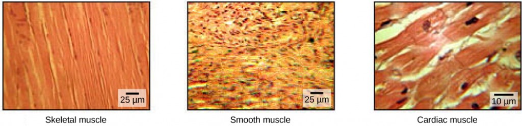
Skeletal muscle tissue forms skeletal muscles, which attach to bones or skin and control locomotion and any movement that tin can exist consciously controlled. Considering information technology can be controlled past thought, skeletal muscle is also called voluntary muscle. Skeletal muscles are long and cylindrical in appearance; when viewed under a microscope, skeletal muscle tissue has a striped or striated appearance. The striations are acquired past the regular arrangement of contractile proteins (actin and myosin). Actin is a globular contractile protein that interacts with myosin for musculus contraction. Skeletal musculus also has multiple nuclei present in a single prison cell.
Smooth muscle tissue occurs in the walls of hollow organs such equally the intestines, stomach, and urinary bladder, and effectually passages such equally the respiratory tract and blood vessels. Shine muscle has no striations, is non nether voluntary control, has simply ane nucleus per prison cell, is tapered at both ends, and is called involuntary musculus.
Cardiac muscle tissue is only found in the heart, and cardiac contractions pump blood throughout the body and maintain blood pressure. Like skeletal musculus, cardiac muscle is striated, but different skeletal musculus, cardiac muscle cannot be consciously controlled and is chosen involuntary muscle. It has 1 nucleus per cell, is branched, and is distinguished by the presence of intercalated disks.
Skeletal Muscle Fiber Structure
Each skeletal muscle fiber is a skeletal musculus cell. These cells are incredibly large, with diameters of up to 100 µm and lengths of upwardly to thirty cm. The plasma membrane of a skeletal muscle fiber is called the sarcolemma. The sarcolemma is the site of action potential conduction, which triggers muscle contraction. Within each muscle fiber are myofibrils—long cylindrical structures that prevarication parallel to the muscle fiber. Myofibrils run the unabridged length of the muscle fiber, and considering they are but approximately 1.2 µm in bore, hundreds to thousands can be found inside one musculus fiber. They adhere to the sarcolemma at their ends, and then that every bit myofibrils shorten, the unabridged muscle cell contracts (Figure 19.34).
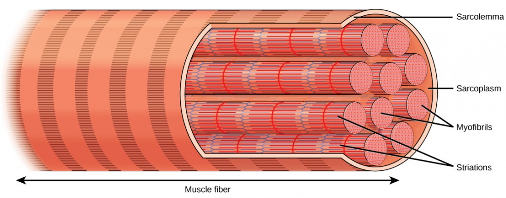
The striated advent of skeletal muscle tissue is a result of repeating bands of the proteins actin and myosin that are present forth the length of myofibrils. Dark A bands and light I bands echo along myofibrils, and the alignment of myofibrils in the jail cell causes the entire cell to appear striated or banded.
Each I ring has a dense line running vertically through the middle called a Z disc or Z line. The Z discs mark the edge of units called sarcomeres, which are the functional units of skeletal muscle. One sarcomere is the infinite between ii consecutive Z discs and contains one unabridged A band and two halves of an I ring, one on either side of the A band. A myofibril is composed of many sarcomeres running along its length, and as the sarcomeres individually contract, the myofibrils and muscle cells shorten (Figure xix.35).
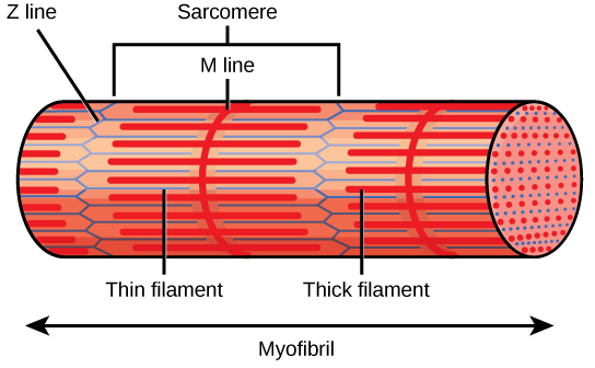
A sarcomere is the region from one Z line to the next Z line. Many sarcomeres are present in a myofibril, resulting in the striation pattern feature of skeletal muscle.
Myofibrils are composed of smaller structures chosen myofilaments. At that place are 2 chief types of filaments: thick filaments and thin filaments; each has different compositions and locations. Thick filaments occur simply in the A band of a myofibril. Sparse filaments attach to a protein in the Z disc called alpha-actinin and occur across the entire length of the I band and partway into the A band. The region at which thick and thin filaments overlap has a dense advent, every bit in that location is little space betwixt the filaments. Thin filaments exercise not extend all the style into the A bands, leaving a central region of the A band that only contains thick filaments. This cardinal region of the A ring looks slightly lighter than the residuum of the A band and is chosen the H zone. The middle of the H zone has a vertical line called the One thousand line, at which accompaniment proteins concord together thick filaments. Both the Z disc and the M line hold myofilaments in place to maintain the structural arrangement and layering of the myofibril. Myofibrils are connected to each other by intermediate, or desmin, filaments that attach to the Z disc.
Thick and thin filaments are themselves composed of proteins. Thick filaments are composed of the protein myosin. The tail of a myosin molecule connects with other myosin molecules to form the central region of a thick filament virtually the K line, whereas the heads marshal on either side of the thick filament where the thin filaments overlap. The principal component of thin filaments is the actin protein. Ii other components of the sparse filament are tropomyosin and troponin. Actin has bounden sites for myosin attachment. Strands of tropomyosin block the binding sites and preclude actin–myosin interactions when the muscles are at residual. Troponin consists of three globular subunits. One subunit binds to tropomyosin, i subunit binds to actin, and one subunit binds Ca2+ ions.
Concept in Action

View this animation showing the organization of muscle fibers.
Sliding Filament Model of Wrinkle
For a muscle cell to contract, the sarcomere must shorten. However, thick and thin filaments—the components of sarcomeres—practice not shorten. Instead, they slide past one another, causing the sarcomere to shorten while the filaments remain the same length. The sliding filament theory of muscle contraction was adult to fit the differences observed in the named bands on the sarcomere at different degrees of muscle contraction and relaxation. The machinery of contraction is the bounden of myosin to actin, forming cantankerous-bridges that generate filament movement (Figure 19.36).
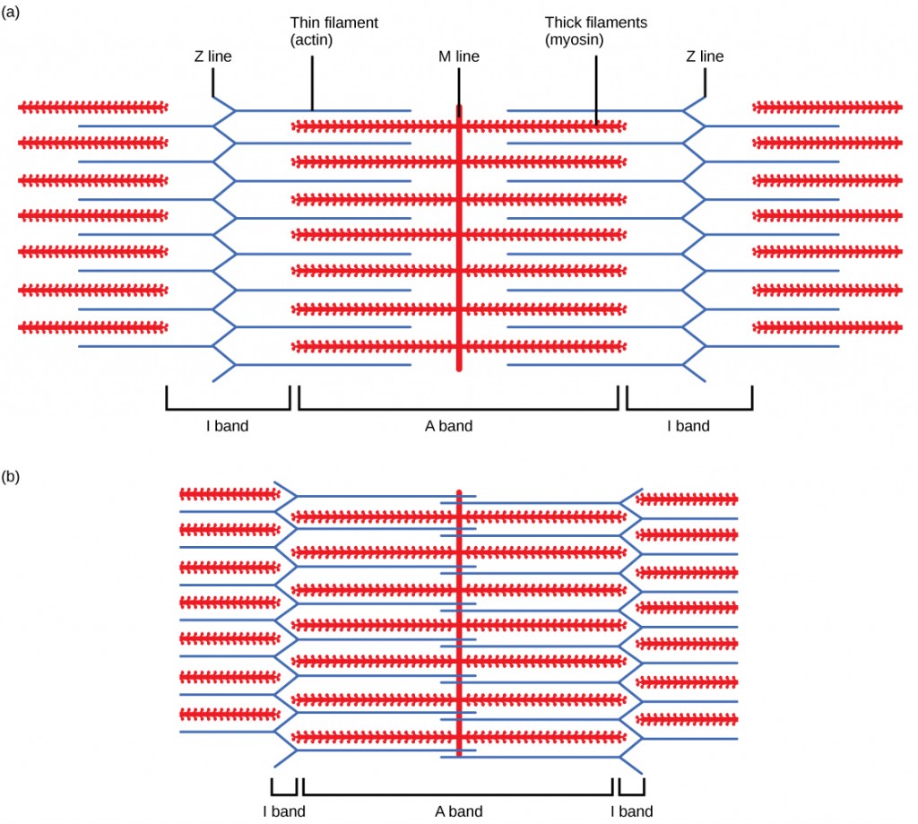
When (a) a sarcomere (b) contracts, the Z lines motion closer together and the I band gets smaller. The A band stays the same width and, at full wrinkle, the sparse filaments overlap.
When a sarcomere shortens, some regions shorten whereas others stay the same length. A sarcomere is defined as the distance between ii consecutive Z discs or Z lines; when a muscle contracts, the distance between the Z discs is reduced. The H zone—the central region of the A zone—contains only thick filaments and is shortened during wrinkle. The I band contains simply thin filaments and too shortens. The A band does not shorten—it remains the same length—merely A bands of unlike sarcomeres movement closer together during wrinkle, eventually disappearing. Thin filaments are pulled by the thick filaments toward the middle of the sarcomere until the Z discs approach the thick filaments. The zone of overlap, in which thin filaments and thick filaments occupy the same area, increases as the thin filaments movement inward.
ATP and Muscle Wrinkle
The motion of musculus shortening occurs every bit myosin heads bind to actin and pull the actin inwards. This action requires energy, which is provided by ATP. Myosin binds to actin at a binding site on the globular actin protein. Myosin has some other binding site for ATP at which enzymatic activity hydrolyzes ATP to ADP, releasing an inorganic phosphate molecule and energy.
ATP binding causes myosin to release actin, assuasive actin and myosin to detach from each other. Later this happens, the newly bound ATP is converted to ADP and inorganic phosphate, Pi. The enzyme at the binding site on myosin is called ATPase. The free energy released during ATP hydrolysis changes the bending of the myosin head into a "cocked" position. The myosin head is and so in a position for further movement, possessing potential energy, but ADP and Pi are even so attached. If actin binding sites are covered and unavailable, the myosin will remain in the high energy configuration with ATP hydrolyzed, but still attached.
If the actin binding sites are uncovered, a cross-span will form; that is, the myosin head spans the distance betwixt the actin and myosin molecules. Pi is then released, allowing myosin to expend the stored energy equally a conformational change. The myosin head moves toward the 1000 line, pulling the actin along with it. As the actin is pulled, the filaments move approximately 10 nm toward the M line. This movement is called the power stroke, as it is the step at which strength is produced. As the actin is pulled toward the M line, the sarcomere shortens and the muscle contracts.
When the myosin head is "artsy," information technology contains free energy and is in a high-free energy configuration. This energy is expended as the myosin head moves through the power stroke; at the end of the power stroke, the myosin head is in a low-energy position. After the power stroke, ADP is released; nonetheless, the cross-bridge formed is nevertheless in place, and actin and myosin are bound together. ATP can and so attach to myosin, which allows the cross-bridge cycle to start once more and further musculus contraction can occur (Figure 19.37).
Concept in Action

Picket this video explaining how a muscle contraction is signaled.
Which of the following statements near musculus contraction is truthful?
- The power stroke occurs when ATP is hydrolyzed to ADP and phosphate.
- The power stroke occurs when ADP and phosphate dissociate from the myosin head.
- The power stroke occurs when ADP and phosphate dissociate from the actin agile site.
- The power stroke occurs when Ca2+ binds the calcium head.
Concept in Activeness

View this animation of the cross-bridge muscle contraction.
Regulatory Proteins
When a muscle is in a resting state, actin and myosin are separated. To proceed actin from binding to the agile site on myosin, regulatory proteins block the molecular binding sites. Tropomyosin blocks myosin binding sites on actin molecules, preventing cross-span formation and preventing wrinkle in a muscle without nervous input. Troponin binds to tropomyosin and helps to position information technology on the actin molecule; it as well binds calcium ions.
To enable a muscle contraction, tropomyosin must change conformation, uncovering the myosin-bounden site on an actin molecule and allowing cantankerous-bridge formation. This can only happen in the presence of calcium, which is kept at extremely low concentrations in the sarcoplasm. If present, calcium ions bind to troponin, causing conformational changes in troponin that allow tropomyosin to move away from the myosin binding sites on actin. In one case the tropomyosin is removed, a cross-span can form betwixt actin and myosin, triggering contraction. Cross-bridge cycling continues until Ca2+ ions and ATP are no longer available and tropomyosin over again covers the binding sites on actin.
Excitation–Contraction Coupling
Excitation–contraction coupling is the link (transduction) betwixt the action potential generated in the sarcolemma and the kickoff of a muscle wrinkle. The trigger for calcium release from the sarcoplasmic reticulum into the sarcoplasm is a neural signal. Each skeletal muscle fiber is controlled by a motor neuron, which conducts signals from the brain or spinal string to the muscle. The area of the sarcolemma on the musculus fiber that interacts with the neuron is chosen the motor cease plate. The finish of the neuron's axon is chosen the synaptic final, and it does not actually contact the motor terminate plate. A small space chosen the synaptic cleft separates the synaptic terminal from the motor terminate plate. Electrical signals travel along the neuron's axon, which branches through the muscle and connects to private muscle fibers at a neuromuscular junction.
The power of cells to communicate electrically requires that the cells expend energy to create an electrical gradient across their cell membranes. This charge gradient is carried by ions, which are differentially distributed across the membrane. Each ion exerts an electrical influence and a concentration influence. Only every bit milk will eventually mix with java without the need to stir, ions as well distribute themselves evenly, if they are permitted to practice then. In this instance, they are not permitted to return to an evenly mixed state.
The sodium–potassium ATPase uses cellular energy to move 1000+ ions inside the prison cell and Na+ ions outside. This alone accumulates a pocket-size electric charge, but a big concentration gradient. There is lots of Chiliad+ in the cell and lots of Na+ exterior the cell. Potassium is able to go out the cell through Thousand+ channels that are open up 90% of the time, and it does. Nonetheless, Na+ channels are rarely open up, and so Na+ remains exterior the cell. When K+ leaves the prison cell, obeying its concentration gradient, that effectively leaves a negative accuse backside. So at rest, at that place is a large concentration slope for Na+ to enter the cell, and there is an aggregating of negative charges left backside in the cell. This is the resting membrane potential. Potential in this context means a separation of electrical charge that is capable of doing work. It is measured in volts, only like a battery. However, the transmembrane potential is considerably smaller (0.07 V); therefore, the pocket-sized value is expressed as millivolts (mV) or 70 mV. Because the within of a cell is negative compared with the exterior, a minus sign signifies the excess of negative charges inside the cell, −70 mV.
If an issue changes the permeability of the membrane to Na+ ions, they will enter the prison cell. That will modify the voltage. This is an electric issue, chosen an action potential, that can exist used as a cellular signal. Communication occurs betwixt fretfulness and muscles through neurotransmitters. Neuron action potentials cause the release of neurotransmitters from the synaptic terminal into the synaptic scissure, where they tin can so diffuse across the synaptic cleft and bind to a receptor molecule on the motor end plate. The motor end plate possesses junctional folds—folds in the sarcolemma that create a large surface area for the neurotransmitter to bind to receptors. The receptors are really sodium channels that open to allow the passage of Na+ into the jail cell when they receive neurotransmitter signal.
Acetylcholine (ACh) is a neurotransmitter released by motor neurons that binds to receptors in the motor end plate. Neurotransmitter release occurs when an action potential travels down the motor neuron's axon, resulting in altered permeability of the synaptic terminal membrane and an influx of calcium. The Catwo+ ions allow synaptic vesicles to motion to and bind with the presynaptic membrane (on the neuron), and release neurotransmitter from the vesicles into the synaptic cleft. Once released by the synaptic last, ACh diffuses across the synaptic crevice to the motor cease plate, where it binds with ACh receptors. As a neurotransmitter binds, these ion channels open, and Na+ ions cross the membrane into the muscle cell. This reduces the voltage difference betwixt the within and outside of the jail cell, which is called depolarization. Every bit ACh binds at the motor finish plate, this depolarization is called an finish-plate potential. The depolarization then spreads along the sarcolemma, creating an action potential as sodium channels adjacent to the initial depolarization site sense the modify in voltage and open. The activeness potential moves across the entire cell, creating a wave of depolarization.
ACh is broken down by the enzyme acetylcholinesterase (Anguish) into acetyl and choline. Ache resides in the synaptic crack, breaking downwards ACh so that information technology does not remain bound to ACh receptors, which would cause unwanted extended muscle contraction (Effigy 19.38).
The mortiferous nerve gas Sarin irreversibly inhibits acetycholinesterase. What upshot would Sarin accept on muscle contraction?
After depolarization, the membrane returns to its resting country. This is called repolarization, during which voltage-gated sodium channels close. Potassium channels continue at 90% conductance. Considering the plasma membrane sodium–potassium ATPase always transports ions, the resting state (negatively charged inside relative to the outside) is restored. The period immediately post-obit the transmission of an impulse in a nerve or muscle, in which a neuron or musculus cell regains its ability to transmit some other impulse, is called the refractory period. During the refractory period, the membrane cannot generate another action potential. . The refractory catamenia allows the voltage-sensitive ion channels to return to their resting configurations. The sodium potassium ATPase continually moves Na+ dorsum out of the cell and M+ back into the cell, and the K+ leaks out leaving negative charge backside. Very chop-chop, the membrane repolarizes, so that it can once more be depolarized.
Control of Muscle Tension
Neural control initiates the formation of actin–myosin cross-bridges, leading to the sarcomere shortening involved in muscle contraction. These contractions extend from the musculus cobweb through connective tissue to pull on basic, causing skeletal movement. The pull exerted by a muscle is chosen tension, and the amount of force created past this tension can vary. This enables the same muscles to move very low-cal objects and very heavy objects. In individual muscle fibers, the amount of tension produced depends on the cross-exclusive area of the musculus fiber and the frequency of neural stimulation.
The number of cross-bridges formed between actin and myosin determine the amount of tension that a muscle fiber can produce. Cross-bridges can only form where thick and sparse filaments overlap, allowing myosin to demark to actin. If more cross-bridges are formed, more than myosin will pull on actin, and more tension will be produced.
The ideal length of a sarcomere during production of maximal tension occurs when thick and sparse filaments overlap to the greatest degree. If a sarcomere at residue is stretched by an ideal resting length, thick and sparse filaments do not overlap to the greatest degree, and fewer cross-bridges can course. This results in fewer myosin heads pulling on actin, and less tension is produced. As a sarcomere is shortened, the zone of overlap is reduced every bit the thin filaments achieve the H zone, which is equanimous of myosin tails. Considering information technology is myosin heads that form cross-bridges, actin will not bind to myosin in this zone, reducing the tension produced by this myofiber. If the sarcomere is shortened fifty-fifty more, thin filaments begin to overlap with each other—reducing cross-bridge formation even further, and producing even less tension. Conversely, if the sarcomere is stretched to the point at which thick and thin filaments exercise not overlap at all, no cross-bridges are formed and no tension is produced. This corporeality of stretching does not normally occur considering accompaniment proteins, internal sensory fretfulness, and connective tissue oppose farthermost stretching.
The primary variable determining strength production is the number of myofibers inside the muscle that receive an action potential from the neuron that controls that fiber. When using the biceps to pick upward a pencil, the motor cortex of the brain only signals a few neurons of the biceps, and only a few myofibers answer. In vertebrates, each myofiber responds fully if stimulated. When picking upwards a piano, the motor cortex signals all of the neurons in the biceps and every myofiber participates. This is close to the maximum force the muscle can produce. As mentioned above, increasing the frequency of action potentials (the number of signals per second) can increase the force a chip more, because the tropomyosin is flooded with calcium.
Summary
The body contains three types of muscle tissue: skeletal muscle, cardiac muscle, and smooth muscle. Skeleton muscle tissue is composed of sarcomeres, the functional units of muscle tissue. Muscle contraction occurs when sarcomeres shorten, every bit thick and thin filaments slide past each other, which is chosen the sliding filament model of muscle wrinkle. ATP provides the energy for cross-bridge formation and filament sliding. Regulatory proteins, such equally troponin and tropomyosin, control cross-span germination. Excitation–contraction coupling transduces the electrical signal of the neuron, via acetylcholine, to an electrical signal on the musculus membrane, which initiates force production. The number of muscle fibers contracting determines how much force the whole muscle produces.
Exercises
- Which of the following statements about muscle contraction is true?
- The power stroke occurs when ATP is hydrolyzed to ADP and phosphate.
- The power stroke occurs when ADP and phosphate dissociate from the myosin head.
- The power stroke occurs when ADP and phosphate dissociate from the actin active site.
- The power stroke occurs when Caii+ binds the calcium head.
- The deadly nerve gas Sarin irreversibly inhibits acetycholinesterase. What consequence would Sarin have on muscle contraction?
- In relaxed musculus, the myosin-bounden site on actin is blocked past ________.
- titin
- troponin
- myglobin
- tropomyosin
- The jail cell membrane of a muscle fiber is called a ________.
- myofibril
- sarcolemma
- sarcoplasm
- myofilament
- The muscle relaxes if no new nervus point arrives. However the neurotransmitter from the previous stimulation is still present in the synapse. The action of ________ helps to remove this neurotransmitter.
- myosin
- action potential
- tropomyosin
- acetylcholinesterase
- The ability of a musculus to generate tension immediately after stimulation is dependent on:
- myosin interaction with the Thou line
- overlap of myosin and actin
- actin attachments to the Z line
- none of the above
- How would muscle contractions be affected if ATP was completely depleted in a muscle fiber?
- What factors contribute to the amount of tension produced in an individual muscle fiber?
- What effect will low blood calcium have on neurons? What effect volition depression claret calcium have on skeletal muscles?
Answers
- B
- In the presence of Sarin, acetycholine is not removed from the synapse, resulting in continuous stimulation of the musculus plasma membrane. At beginning, muscle activity is intense and uncontrolled, merely the ion gradients dissipate, so electrical signals in the T-tubules are no longer possible. The result is paralysis, leading to decease by asphyxiation.
- D
- B
- D
- D
- Considering ATP is required for myosin to release from actin, muscles would remain rigidly contracted until more ATP was available for the myosin cross-bridge release. This is why dead vertebrates undergo rigor mortis.
- The cross-exclusive area, the length of the muscle fiber at residuum, and the frequency of neural stimulation.
- Neurons volition not exist able to release neurotransmitter without calcium. Skeletal muscles have calcium stored and don't demand any from the exterior.
Glossary
- acetylcholinesterase
- (Ache) enzyme that breaks down ACh into acetyl and choline
- actin
- globular contractile protein that interacts with myosin for muscle contraction
- motor stop plate
- sarcolemma of the muscle fiber that interacts with the neuron
- myofibril
- long cylindrical structures that lie parallel to the musculus fiber
- myofilament
- modest structures that make up myofibrils
- myosin
- contractile protein that interacts with actin for muscle contraction
- osseous tissue
- connective tissue that constitutes the endoskeleton
- sarcolemma
- plasma membrane of a skeletal musculus fiber
- sarcomere
- functional unit of skeletal musculus
- skeletal muscle tissue
- forms skeletal muscles, which attach to bones and control locomotion and whatsoever movement that can be consciously controlled
- spongy bone tissue
- forms the inner layer of all bones
- thick filament
- a grouping of myosin molecules
- sparse filament
- two polymers of actin wound together along with tropomyosin and troponin
- tropomyosin
- acts to block myosin binding sites on actin molecules, preventing cross-bridge formation and preventing contraction until a muscle receives a neuron signal
- troponin
- binds to tropomyosin and helps to position it on the actin molecule, and as well binds calcium ions
- ulna
- bone located on the medial aspect (pinky-finger side) of the forearm
- vertebral column
- (also, spine) surrounds and protects the spinal cord, supports the head, and acts as an attachment point for ribs and muscles of the back and cervix
Source: https://opentextbc.ca/biology/chapter/19-4-muscle-contraction-and-locomotion/
Post a Comment for "Review Another Set of Terms Associated With a Skeletal Muscle Fiber/cell and the Sarcomere"